Recent improvements in AFM imaging modes and technology are providing an unprecedented level of control for imaging biomolecules in situ at high resolution. The stiffness and adhesion of cells are providing vital disease indicators.
Cells and biomolecules are at home in a liquid environment of well defined composition and temperature. AFM is suitable for liquid phase measurements, and can often provide unique insights on in situ behaviour. Relevant measurements include topography and property maps where the effect of additives on cell morphology and stiffness can be observed, and force spectroscopy for a more detailed analysis of cell mechanics. At the molecular level, the conformation and e.g. hybridisation of DNA, peptides, proteins, lipids and biomembranes can be investigated.
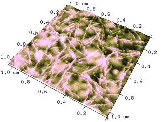
Spider Silk Peptides on Gold
Two scales of features are observable - the molecular bead-and-necklace structure of recombinant spider silk adsorbed onto the characteristic structure of evaporated gold. The image was obtained in buffer solution with Peak Force Tapping on the Bruker Dimension FastScan Bio system.
Sample courtesy of Linnea Nilebäck and My Hedhammar, School of Biotechnology, KTH
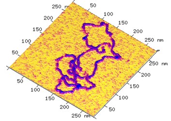
Liquid-phase Imaging of DNA
Plasmid DNA immobilized on mica in Ni2+ containing HEPES buffer. Image taken in Peak Force Tapping mode in buffer on the Bruker Dimension FastScan Bio system.
Samples courtesy of Andris Abramenkovs and Bo Stenerlöw, Uppsala University
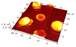
Imaging of Cells
Human red blood cells infected with malaria parasite. The cells are attached to a glass slide with polylysine and imaged in air in Tapping Mode with the Bruker Dimension FastScan system, scan-rate 2 kHz.
Sample courtesy of Junhong Ch’ng and Mats Wahlgren, Karolinska Institutet
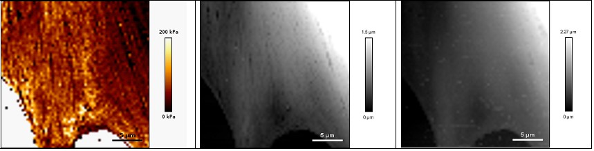
The mechanical properties of living cells play an important role in their function. Here, a sharp imaging tip is used to indent the cell in a high resolution matrix that after analysis provides maps of the stiffness and height of the cell (Quantitative Imaging (or QI) Mode, JPK Instruments). Here, Young's Modulus, which is a measure of stiffness, is mapped over a fibroblast cell in medium at 37oC. The height at maximum force ("inside" the cell) and the surface profile are also displayed. The effect of drugs and other additives were then investigated.
Samples courtesy of Annica Gad, Karolinska Institutet